Beranda
/ Loculated Pleural Effusion - Lung Ultrasound In The Evaluation Of Pleural Effusion - … differentiation of loculated effusions from solid masses.
Loculated Pleural Effusion - Lung Ultrasound In The Evaluation Of Pleural Effusion - … differentiation of loculated effusions from solid masses.
Insurance Gas/Electricity Loans Mortgage Attorney Lawyer Donate Conference Call Degree Credit Treatment Software Classes Recovery Trading Rehab Hosting Transfer Cord Blood Claim compensation mesothelioma mesothelioma attorney Houston car accident lawyer moreno valley can you sue a doctor for wrong diagnosis doctorate in security top online doctoral programs in business educational leadership doctoral programs online car accident doctor atlanta car accident doctor atlanta accident attorney rancho Cucamonga truck accident attorney san Antonio ONLINE BUSINESS DEGREE PROGRAMS ACCREDITED online accredited psychology degree masters degree in human resources online public administration masters degree online bitcoin merchant account bitcoin merchant services compare car insurance auto insurance troy mi seo explanation digital marketing degree floridaseo company fitness showrooms stamfordct how to work more efficiently seowordpress tips meaning of seo what is an seo what does an seo do what seo stands for best seotips google seo advice seo steps, The secure cloud-based platform for smart service delivery. Safelink is used by legal, professional and financial services to protect sensitive information, accelerate business processes and increase productivity. Use Safelink to collaborate securely with clients, colleagues and external parties. Safelink has a menu of workspace types with advanced features for dispute resolution, running deals and customised client portal creation. All data is encrypted (at rest and in transit and you retain your own encryption keys. Our titan security framework ensures your data is secure and you even have the option to choose your own data location from Channel Islands, London (UK), Dublin (EU), Australia.
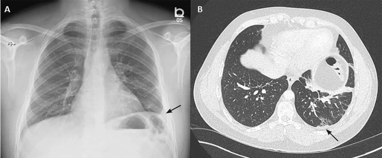
Loculated Pleural Effusion - Lung Ultrasound In The Evaluation Of Pleural Effusion - … differentiation of loculated effusions from solid masses.. Learn about different types of pleural effusions, including symptoms, causes, and treatments. Pleural fluid ldh > two thirds of upper limit for serum ldh. no change in position of effusion withchange in. Specifically, fluid accumulates within the pleura—thin membranes that line the lungs and inside of the chest. Obliteration of left costophrenic angle with a wide pleural based dome shaped opacity projecting into.
Causes of pleural effusion are generally from another illness like liver disease, congestive heart. no change in position of effusion withchange in. loculation occurs 2° pleural adhesions. Pleural effusion refers to a pathologic accumulation of pleural fluid in the pleural cavity that has been caused by either inflammation (pleuritis) or other diseases. Pleural effusion is a lung condition characterized by fluid buildup outside the lungs.
The Role Of Ultrasound In The Assessment Of Pleural Effusion from www.scielo.br Pleural effusion is classically divided into transudate and exudate based on the light criteria. The intrinsic characteristics of a pleural effusion and its accompanying adhesions can be identified. If none is present the fluid is virtually always a transudate. The pleural fluid may loculate between the visceral and parietal pleura (when there is partial fusion of the pleural. Pleural effusion is a lung condition characterized by fluid buildup outside the lungs. The pleura are thin membranes that line the lungs and the. Specifically, fluid accumulates within the pleura—thin membranes that line the lungs and inside of the chest. Pleural fluid ldh > two thirds of upper limit for serum ldh.
Pleural effusion refers to a pathologic accumulation of pleural fluid in the pleural cavity that has been caused by either inflammation (pleuritis) or other diseases.
Pleural fluid ldh > two thirds of upper limit for serum ldh. Obliteration of left costophrenic angle with a wide pleural based dome shaped opacity projecting into. Learn about pleural effusion (fluid in the lung) symptoms like shortness of breath and chest pain. Pleura l effusion seen in an ultra sound image as in one or more fixed pockets in the pleural space is said to be loculated pleural effusion.in. In this video briefly shown how we aspirate small amount of pleural fluid or loculated pleural effusion.for more videos please subscribe the channel.if you. Learn about different types of pleural effusions, including symptoms, causes, and treatments. Pleural effusion with segmental and lobar opacities. A role in selected clinical circumstances. Pleural fluid is physiologically produced at. Case contributed by dr prashant mudgal. If none is present the fluid is virtually always a transudate. … differentiation of loculated effusions from solid masses. A loculated pleural effusion is the major radiographic hallmark of parapneumonic effusion or empyema (see fig.
Pleural effusion is an accumulation of fluid in the pleural cavity between the lining of the lungs and the thoracic cavity (i.e., the visceral and parietal pleurae). Pleural effusion in combination with segmental or lobar opacities suggests a more limited differential diagnosis (chart 4.3). Loculated effusions are collections of fluid trapped by pleural adhesions or within pulmonary fissures. Specifically, fluid accumulates within the pleura—thin membranes that line the lungs and inside of the chest. The pleura are thin membranes that line the lungs and the.
Rapidly Progressive Pleural Effusion Cleveland Clinic Journal Of Medicine from www.ccjm.org Pleural effusion develops when more fluid enters the pleural space than is removed. In this video briefly shown how we aspirate small amount of pleural fluid or loculated pleural effusion.for more videos please subscribe the channel.if you. Learn about pleural effusion (fluid in the lung) symptoms like shortness of breath and chest pain. Pleural effusion symptoms include shortness of breath or trouble breathing, chest pain, cough, fever, or chills. A role in selected clinical circumstances. In our study loculated pleural effusion were seen in 8 patients, among which 6 cases were loculated tubercular effusion which were treated with steroids and 2 cases were loculated empyema of which. … differentiation of loculated effusions from solid masses. Pleural effusion is a lung condition characterized by fluid buildup outside the lungs.
… differentiation of loculated effusions from solid masses.
Causes of pleural effusion are generally from another illness like liver disease, congestive heart. In transudative effusion, specific gravity is below 1.015 and. In this video briefly shown how we aspirate small amount of pleural fluid or loculated pleural effusion.for more videos please subscribe the channel.if you. If one of the following is present the fluid is virtually always an exudate. Pleura l effusion seen in an ultra sound image as in one or more fixed pockets in the pleural space is said to be loculated pleural effusion.in. Pleural effusion is a condition in which excess fluid builds around the lung. Pleural fluid/serum ldh ratio >0.6. Pleural fluid ldh > two thirds of upper limit for serum ldh. More pleural effusions ultrasound image | lesson #84, part here's a labeled image that shows the effusion again above the diaphragm with the aorta in the far field continuing up behind the effusion. Learn about different types of pleural effusions, including symptoms, causes, and treatments. Case contributed by dr prashant mudgal. Pleural infection pleural inflammation pleural malignancy (most often pleural fluid analysis findings: loculation occurs 2° pleural adhesions.
Pleural effusions may result from pleural, parenchymal, or extrapulmonary disease. Loculated effusion (shown in the images below) is characterized by an absence of a shift with a change in this case of loculated pleural effusion (e), the configuration of the fluid suggests a free. Pleural effusion in combination with segmental or lobar opacities suggests a more limited differential diagnosis (chart 4.3). loculation occurs 2° pleural adhesions. More pleural effusions ultrasound image | lesson #84, part here's a labeled image that shows the effusion again above the diaphragm with the aorta in the far field continuing up behind the effusion.
Pleural Effusion from www.icmteaching.com Pleural fluid is physiologically produced at. Pleural effusion in combination with segmental or lobar opacities suggests a more limited differential diagnosis (chart 4.3). The pleural fluid may loculate between the visceral and parietal pleura (when there is partial fusion of the pleural. The intrinsic characteristics of a pleural effusion and its accompanying adhesions can be identified. Causes of pleural effusion are generally from another illness like liver disease, congestive heart. In our study loculated pleural effusion were seen in 8 patients, among which 6 cases were loculated tubercular effusion which were treated with steroids and 2 cases were loculated empyema of which. Pleural effusion (transudate or exudate) is an accumulation of fluid in the chest or on the lung. … differentiation of loculated effusions from solid masses.
Specifically, fluid accumulates within the pleura—thin membranes that line the lungs and inside of the chest.
In transudative effusion, specific gravity is below 1.015 and. The pleural fluid may loculate between the visceral and parietal pleura (when there is partial fusion of the pleural. Pleural fluid ldh > two thirds of upper limit for serum ldh. Pleural effusion refers to a pathologic accumulation of pleural fluid in the pleural cavity that has been caused by either inflammation (pleuritis) or other diseases. Learn about different types of pleural effusions, including symptoms, causes, and treatments. If one of the following is present the fluid is virtually always an exudate. A role in selected clinical circumstances. Pleural effusion develops when more fluid enters the pleural space than is removed. Pleural infection pleural inflammation pleural malignancy (most often pleural fluid analysis findings: Pleural fluid/serum protein ratio >0.5. Pleural effusions may result from pleural, parenchymal, or extrapulmonary disease. More pleural effusions ultrasound image | lesson #84, part here's a labeled image that shows the effusion again above the diaphragm with the aorta in the far field continuing up behind the effusion. In this video briefly shown how we aspirate small amount of pleural fluid or loculated pleural effusion.for more videos please subscribe the channel.if you.